Eric A. Brandser, M.D.
Peer Review Status: Internally Peer Reviewed
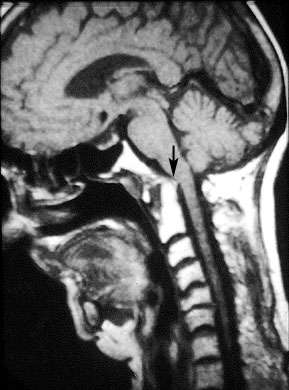
The vertebral bodies appear white due to the large amount of fat located in the marrow. Notice that the gap between C1 and C2 is too wide. There is a synovial joint that sits between the odontoid process and anterior arch of C1 which can be affected by RA. As the pannus erodes the bone, it will also degenerate the alar check ligaments which stablize C1. If the ligaments are weakened then C1 can impinge upon the spinal cord (arrow). Injury to the cord occasionally occurs in surgery when the head is tilted back to insert the endotracheal tube. This is why anesthesia will occasionally have orthopedics check for C1-C2 laxity in surgical patients with RA. The patient is asked to flex and extend their neck and if they display a large degree of motion, then a CT scan is generated to check the vertebrae.
Return to the Radiological Arthritis Evidence Entrance
Copyright protected material used with permission of the author and the University of Iowa’s Virtual Hospital, www.vh.org